3C. T Cell Overview
Origin, Maturation and Antigen Recognition
T cells mature and undergo positive and negative selection processes in the thymus (determining if T cell receptors bind to body's MHC, if binding too weakly, they are 'neglected' and undergo apoptosis).
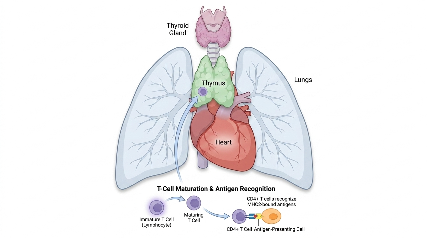
T Cells are vital in both components of active immunity, including cell mediated immunity and to some extent, humoral. The most commonly known T cells are CD4+ T cells (helper T), and CD8+ T cells (cytotoxic/killer T).
T cells cannot recognise soluble free antigens, only receptor bound, protein based antigens. Recognition occurs via MHC (also known as HLA) 1 & 2 receptors along with T cell receptors (TCR) which bind the antigen and form a complex that allows for recognition.
CD4+ T cells recognise MHC2 bound antigens, whilst CD8+ T cells recognise MHC1 bound antigens. T cells also carry a pan-T cell co receptor, CD3.
CD4+ T Cells
CD4+ T cells are known as T Helper cells, and they recognise antigens bound to MHC class II molecules. CD4+ cells are effector cells that produce cytokines to activate other immune cells. They also activate the T-cell-dependent branch of humoral immunity, helping B cells to produce antibodies.
CD8+ T Cells
CD8+ T cells are known as cytotoxic, or killer T cells. They recognise antigens bound to MHC class I molecules. Once activated, they kill target cells (i.e. virally infected cells) through two pathways:
- Fas/FasL pathway; triggering apoptosis via caspase activation
- Release of granzymes and perforin; compounds that bypass the cell membrane and activate caspases directly.
CD4+ Helper Subtypes
| Subtype | Key Cytokines | Primary Role |
|---|---|---|
| Th1 | IFN- | Activates macrophages; fights intracellular infections; implicated in delayed-type hypersensitivity DTH |
| Th2 | IL-4, IL-5, IL-13 | Combats helminthic (parasitic) infections; activates mast cells & eosinophils; involved in allergies |
| Th17 | IL-17A, IL-17F, IL-22 | Mucosal immunity against extracellular bacteria and fungi; drives inflammation; implicated in autoimmune disorders such as IBD and rheumatoid arthritis |
T regs (regulatory T cells), and Tfh cells (T follicular helper cells), are also CD4+ T helper subtypes.
Regulatory T Cells
Regulatory T cells are commonly referred to as T regs, and also known as suppressor T cells. They modulate immune response, and inhibit autoimmunity. They produce inhibitory cytokines including IL-10 and IL-35. They use CTLA-4 (Cytotoxic T-lymphocyte-associated protein 4), to supress antigen presenting cells. Their development is driven by IL-2 and TGF-
Gamma-Delta T cells
A small subpopulation of T cells, featuring T cell receptors made of gamma and delta chains instead of the usual alpha and delta chains. They recognise different antigen types and reside mostly in mucosal and epithelial layers.
T Cell Clinical Relevance
- HIV targets CD4+ Helper T cells via CCR5 and CYCR4 chemokine receptors.
- HTLV (human T cells lymphotropic virus) also targets CD4+ T helper cells and can lead to adult T cell leukaemia and lymphoma.
- In DiGeorge syndrome the thymus never forms so T cells never mature.
- In Bare Lymphocyte Syndrome, impaired MHC expression leads to T cell loss.
- T cells are central to transplant rejection and are pharmacologic targets for anti-rejection drugs
Histology
T cells are identified by flow cytometry using their CD markers, and not by light or electron microscopy. Although, under electron microscopy they have a smoother surface and have more electron dense cytoplasm compared to B cells.
Source
Source: Sauls, R. S., McCausland, C., & Taylor, B. N. (2023, May 1). Histology, T-Cell Lymphocyte. Retrieved from PubMed website: https://www.ncbi.nlm.nih.gov/books/NBK535433/
next page | back to contents